
Knee Treatment & Robotic Knee Replacement in Nashik
From conservative arthritis management and keyhole arthroscopy to subvastus muscle-sparing robotic total and partial knee replacement. Evaluated and operated by Dr. Vishal Kasliwal at Kasliwal Joint Institute.
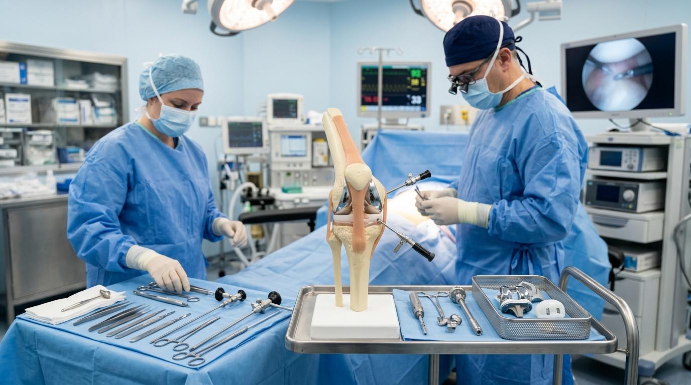
Common Knee Problems We Treat
Every knee problem has a distinct anatomical origin. Accurate clinical diagnosis with weight-bearing imaging ensures targeted, conservative-first care.
Knee Osteoarthritis
Progressive thinning of joint cartilage causing friction, bone spurs, stiffness, and bowing deformity (varus knees).
Ligament Tears (ACL, PCL)
Sudden pivoting sports injuries or two-wheeler trauma causing joint instability, giving way, and recurrent swelling.
Meniscus Tears
Torn shock-absorbing knee cartilage resulting in joint-line tenderness, painful clicking, catching, and difficulty squatting.
Chondral Defects & Loose Bodies
Isolated areas of damaged articular cartilage or loose joint fragments causing painful knee locking and early wear.
Patella Instability & Maltracking
The kneecap slipping out of its femoral groove, leading to recurrent dislocations, anterior pain, and apprehension during stairs.
Painful or Loose Replacement
Persistent pain, instability, or implant loosening from a prior surgery requiring specialist revision assessment.
Knee Arthritis Treatment in Nashik
Knee osteoarthritis is the progressive wear of the smooth cartilage covering the ends of the femur and tibia. In Indian patients, wear frequently concentrates on the medial (inner) side, resulting in gradual bow-legged alignment.
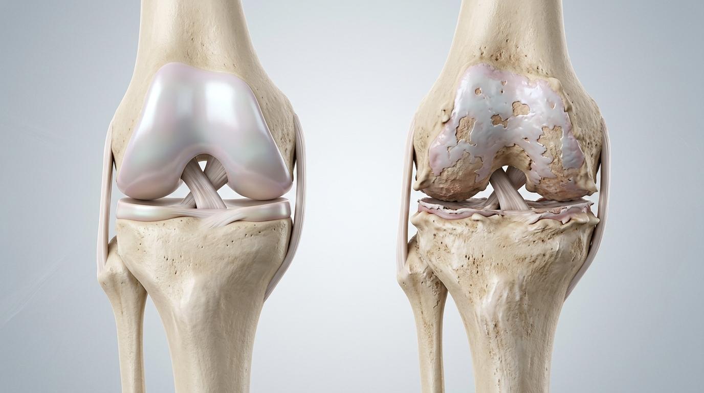
Types of Knee Arthritis
- •Primary Osteoarthritis: The most common form, related to age, biomechanical wear, genetic factors, and weight.
- •Inflammatory Arthritis: Systemic autoimmune conditions such as Rheumatoid Arthritis affecting both knee joints.
- •Post-Traumatic Arthritis: Secondary cartilage degeneration occurring years after a previous fracture, ACL rupture, or meniscus tear.
Key Warning Signs & Symptoms
Accurate Standing X-Ray & Alignment Scanograms
We perform full weight-bearing standing views and digital limb scanograms to evaluate true mechanical load axes and joint space narrowing before planning treatment.
The 6-Step Non-Surgical Treatment Ladder
Surgery is never the initial step. For early to moderate arthritis, evidence-based non-surgical therapies control inflammation, strengthen supporting muscles, and preserve joint function.
Physiotherapy & Muscle Strengthening
Focused isometric quadriceps, vastus medialis, and hamstring training creates a muscular shock absorber around the joint.
Weight Management
Every 1 kg of body weight lost relieves approximately 4 kg of cumulative compressive stress across the knee joint with each step.
Activity Modification
Transitioning from high-impact activities to cycling, swimming, and controlled flat-surface walking; avoiding deep squatting.
Monitored Medical Therapy
Short, monitored courses of anti-inflammatory medications and cartilage protectants to quiet acute inflammatory flare-ups.
Unloading Knee Braces
Custom off-loader knee braces transfer mechanical load away from the worn inner compartment onto healthy lateral cartilage.
Targeted Joint Injections
Hyaluronic acid viscosupplementation or targeted anti-inflammatory injections for patients needing interim relief.
Do You Need a Knee Replacement?
Surgery is indicated when arthritis pain interferes with independent daily life and conservative measures stop working. Review these 7 clinical indicators:
If you answered yes to 3 or more indicators, an objective surgical consultation is recommended.
Book Knee EvaluationVELYS™ Robotic Total Knee Replacement (TKR)
In a total knee replacement, the worn cartilage surfaces of the thigh bone (femur) and shin bone (tibia) are resurfaced with precision cobalt-chromium or titanium components, cushioned by a high-durability polyethylene insert.
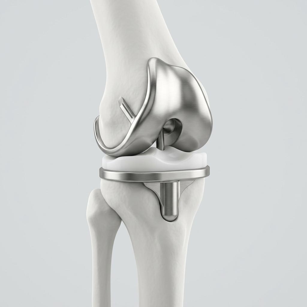
Complete Kinematic Joint Resurfacing System
Subvastus Muscle-Sparing Surgical Approach
Dr. Kasliwal utilizes a muscle-sparing subvastus approach whenever anatomically suitable. By elevating rather than cutting the quadriceps tendon, natural muscle control is retained, resulting in reduced postoperative pain and faster independent leg lifting.
Personalised Kinematic Alignment
Rather than forcing a rigid 90-degree mechanical angle for every patient, VELYS™ robotic assistance maps your unique natural joint line and soft-tissue balance, restoring the natural feel of your knee.
Real-Time Ligament Balancing Across Full Range
The robotic optical tracking array measures joint tension dynamically through 0° extension to 120° flexion, ensuring stable, balanced knee movement without tight spots or instability.
Partial Knee Replacement (Unicompartmental UKR)
Not every arthritic knee needs a total replacement. When wear is confined to the medial (inner) compartment, replacing only that single damaged compartment preserves the remaining 70% of healthy bone and both crucial cruciate ligaments (ACL and PCL).
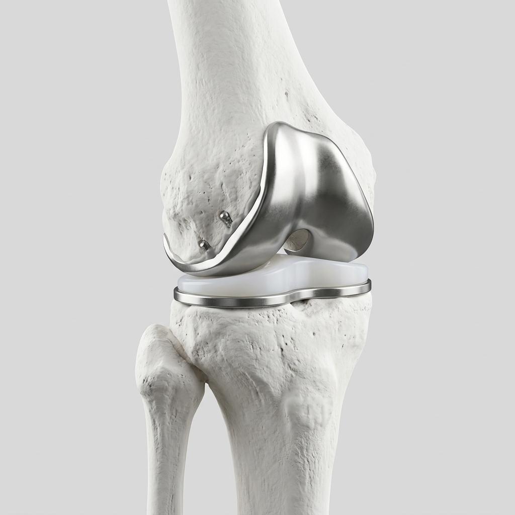
Unicompartmental Bone-Preserving System
| Clinical Feature | Partial Knee (UKR) | Total Knee (TKR) |
|---|---|---|
| Area Replaced | Single compartment (medial/lateral) | All 3 compartments resurfaced |
| Ligaments Preserved | Both ACL & PCL intact | Substituted by implant design |
| Incision Length | Smaller (approx. 7–8 cm) | Standard (approx. 10–13 cm) |
| Natural Joint Feel | Closer to normal knee proprioception | Stable, predictable, pain-free feel |
| Suitability | Isolated unicompartmental arthritis | Advanced multi-compartment wear |
Recovery Timeline After Knee Replacement
With muscle-sparing subvastus techniques and targeted nerve blocks, recovery follows a predictable, structured timeline:
Standing & First Steps
Supervised standing and walking with a walker occurs within 24 hours. Early quadriceps isometric sets and ankle pumps begin.
Corridor & Stair Practice
Bed-to-chair independence, knee flexion past 90 degrees, and step/stair training with our physiotherapy team before discharge.
Wound Review & Stick Walking
Surgical dressing review; transition from walker to single walking stick. Independent home mobility for basic routines.
Independent Walking
Most patients walk comfortably without any assistive stick. Resumption of light household activities and outdoor walks.
Driving & Office Work
Return to desk work, domestic travel, and driving (once emergency brake reaction time is safe and approved by surgeon).
Full Strength & Endurance
Continued gains in quadriceps endurance, stair climbing ease, and natural joint feeling as residual deep tissue swelling subsides.
Indian Lifestyle & Home Setup Guidance
Standard chair seating, dining table usage, and stair navigation are fully resumed. While high-flexion implants technically allow cross-legged sitting for some patients, deep squatting and using ground-level Indian toilets exert extreme peak forces across prosthetic components. A Western raised commode seat and chair seating are strongly advised to maximize the 15–20+ year lifespan of your implant.
Surgical Safety & Risk Mitigation
Knee replacement is one of modern medicine's most successful operations. We manage and mitigate known surgical risks through strict clinical protocols:
Infection Prevention Protocols
Surgeries are performed inside ultra-clean modular operating theatres equipped with laminar airflow and HEPA filtration. Patients receive weight-adjusted preoperative antibiotic prophylaxis adhering strictly to ICMR orthopaedic safety standards.
Blood Clot (DVT) Prevention
Deep vein thrombosis risks are minimized with multimodal thromboprophylaxis: immediate Day 1 mobilisation, intermittent pneumatic calf compression pumps, and prophylactic blood-thinning medications.
Joint Stiffness & Scar Management
Early active-assisted knee flexion within 24 hours prevents arthrofibrosis. Personalized physiotherapy protocols guide patients to achieve 110° to 125° comfortable bending.
Implant Longevity & Wear Resistance
Sub-millimeter robotic bone preparation and kinematic ligament balance ensure that joint load is evenly distributed across bearing surfaces, preventing premature edge-loading and polyethylene wear.
Knee Replacement Cost & Cashless Insurance in Nashik
Every surgical estimate is provided in writing following your clinical consultation and standing X-ray review. The total cost is determined by four clinical factors:
Procedure Type
Single knee vs bilateral same-day; partial (UKR) vs total robotic replacement.
Implant System
Implant design, bearing surface specifications, and constraint requirements.
Room Category
Choice of general healthcare ward, semi-private room, or private deluxe suite.
Stay Duration
Standard 3–4 day stay or extended medical observation for co-morbid conditions.
Cashless Mediclaim Desk & TPA Empanelment
Our in-house insurance desk manages pre-authorization and claims with all major public and private health insurance companies. We help verify your coverage, policy waiting periods, and room eligibility prior to admission.
Frequently Asked Questions About Knee Care
Considering Knee Replacement or Arthritis Care?
Schedule an in-person assessment with Dr. Vishal Kasliwal to evaluate your knee joints, review your standing X-rays, and understand your personalized treatment ladder.
Medically reviewed by: Dr. Vishal Kasliwal, M.S. Orthopaedics · Reg. No. MMC/2005/02/1098
Last updated: October 2026
Medical Disclaimer: This information is for educational purposes only and does not replace an individual medical consultation. Diagnosis, treatment options, and recovery timelines differ between patients.