Knee & Shoulder Arthroscopy in Nashik
Advanced keyhole surgery for ACL tears, meniscus injuries, joint cartilage restoration, and shoulder rotator cuff repairs, backed by 24/7 emergency fracture and orthopaedic trauma care. Fellowship-trained expertise with Dr. Vishal Kasliwal at Kasliwal Joint Institute.
What is Arthroscopy?
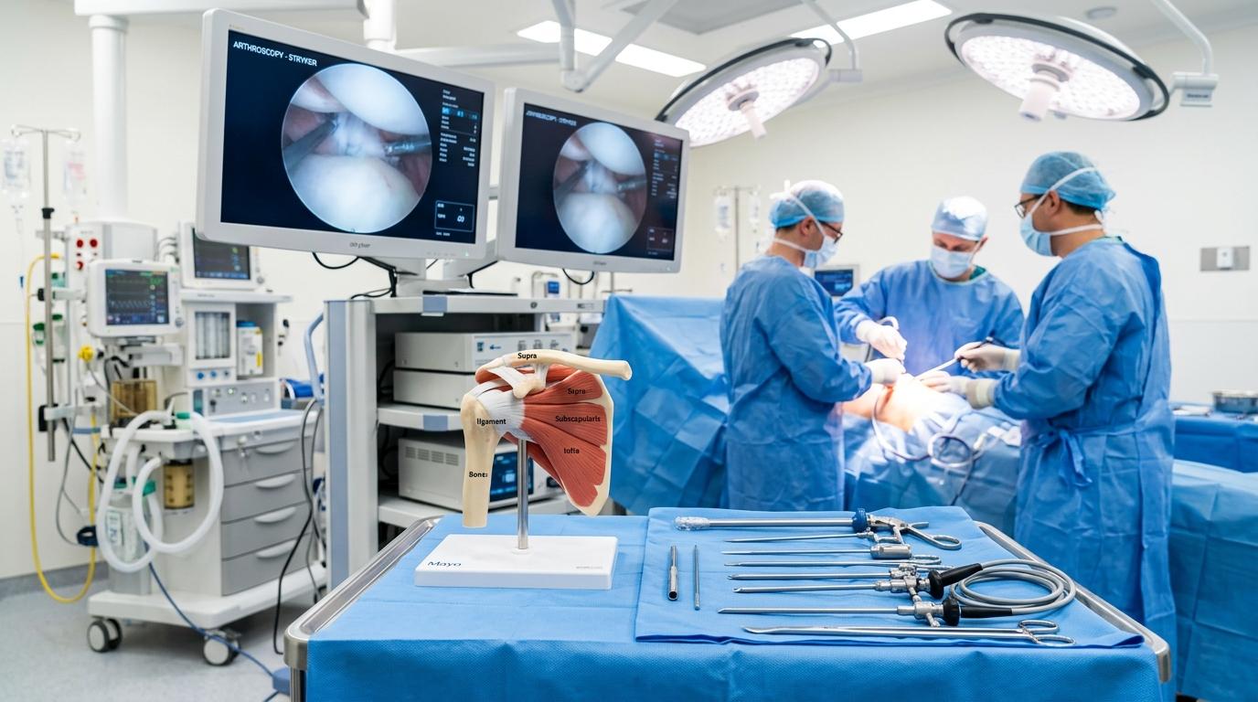
Arthroscopy is a state-of-the-art keyhole technique where a slender optical telescope (arthroscope) and precision micro-instruments are guided into the joint cavity through tiny incisions. High-definition magnification allows meticulous reconstruction of torn ligaments and cartilage without cutting open muscles.
4–5 mm Keyhole Portals
Instead of wide open joint exposures, procedures are completed through puncture incisions, dramatically reducing post-operative pain, scar tissue, and infection risks.
4K High-Definition Visualization
Ultra-high-definition optics illuminate hidden joint recesses, allowing exact probing, tensioning, and anatomical suture fixation of damaged tissue under direct view.
Rapid Day-Care Recovery
Most patients mobilize on the same day and are discharged within 24 hours. Physiotherapy commences immediately, returning patients to desk work and daily routines swiftly.
ACL Tear & Arthroscopic ACL Reconstruction
The Anterior Cruciate Ligament (ACL) is the primary internal stabilizer preventing the tibia from sliding forward or rotating erratically. ACL ruptures occur during rapid cutting, pivoting, awkward jump landings, or two-wheeler traffic collisions.
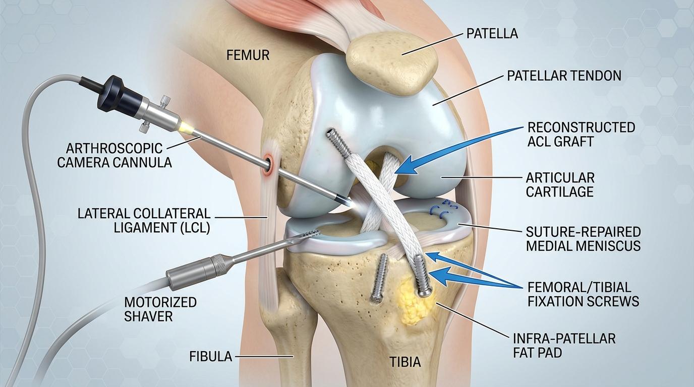
3D Anatomical Illustration: Portal camera, ACL reconstruction graft & meniscus repair.
Who Requires Reconstruction Surgery?
While non-operative rehabilitation may manage sedentary patients with stable knees, active individuals and athletes require anatomical reconstruction. Without an intact ACL, recurrent knee shifts cause secondary meniscus tears and accelerate degenerative joint wear.
Anatomical Autograft Reconstruction
Dr. Vishal Kasliwal reconstructs the torn ligament using a living autograft (quadrupled hamstring or patellar tendon) harvested via a tiny cosmetic incision. Rigid titanium suspensory buttons and bio-absorbable interference screws ensure robust fixation and early mobilization.
Meniscus Tear Treatment: Repair vs. Trimming
The menisci are two C-shaped fibrocartilaginous shock absorbers distributing loads between the femur and tibia. In young athletes, tears occur from sudden rotational pivots; in older adults, tears can develop from minor twisting on degenerated cartilage.
| Clinical Aspect | Meniscus Repair (Preservation) | Partial Meniscectomy (Trimming) |
|---|---|---|
| Surgical Approach | Torn cartilage margins are stitched back together using all-inside micro-sutures. | Only the unstable, torn flap that lacks healing capacity is smoothly resected. |
| Ideal Candidates | Vascular outer edge ("red-red" zone) tears, young athletes, and meniscus root tears. | Avascular inner edge ("white-white" zone) tears and complex degenerative flaps. |
| Rehabilitation Timeline | Protected brace weight-bearing for 4 to 6 weeks to allow biological healing. | Rapid recovery with early weight-bearing and return to normal walking within 1 to 2 weeks. |
| Long-Term Benefit | Preserves native joint shock absorption, lowering long-term risk of early arthritis. | Immediate relief from painful joint locking, catching, and mechanical irritation. |
PCL, Cartilage Restoration & Kneecap Instability
Beyond isolated ACL and meniscus tears, high-energy impact and developmental anatomy can destabilize other structural stabilizers of the knee joint.
PCL & Multi-Ligament Tears
The posterior cruciate ligament (PCL) is damaged during dashboard impacts in car crashes or heavy falls onto a bent knee. Isolated tears are managed with specialized dynamic bracing, whereas multi-ligament instability requires arthroscopic reconstruction to avoid chronic posterior subluxation.
Cartilage Restoration Procedures
Localized cartilage defects cause persistent swelling, sharp pain, and mechanical clicking. Depending on defect depth, interventions range from arthroscopic microfracture (stimulating stem cell marrow release) to osteochondral autograft transfer (OATS) to resurface weight-bearing zones.
Kneecap (MPFL) Reconstruction
Recurrent patellar dislocation occurs when the kneecap slips outward from the femoral groove during pivoting, commonly in adolescents. Surgical reconstruction of the Medial Patellofemoral Ligament (MPFL) restores anatomical alignment and prevents future dislocation episodes.
Shoulder Arthroscopy in Nashik
The shoulder is the most mobile joint in the human body, relying on delicate soft-tissue restraints for stability. Keyhole shoulder surgery repairs complex tears with minimal stiffness and tissue trauma.
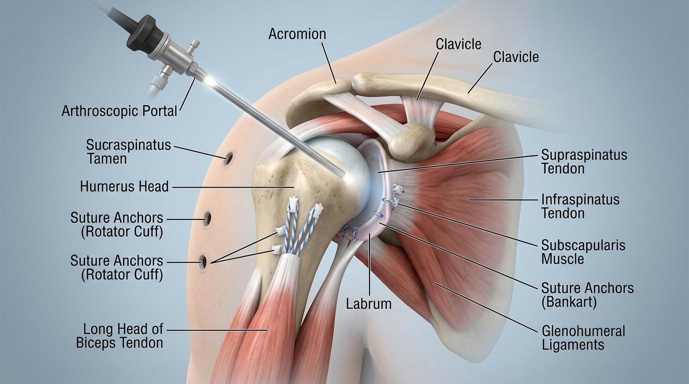
3D Anatomical Illustration: Rotator cuff tendon repair with suture anchors & Bankart labral refixation.
Rotator Cuff Tear Repair
Tears in the supraspinatus tendon cause severe night ache and arm lifting weakness. Symptomatic tears that fail conservative physiotherapy are re-anchored onto the humeral bone footprint using bio-composite suture anchors via 3 tiny keyhole portals, restoring shoulder strength and active range of motion.
Shoulder Dislocation & Bankart Repair
Traumatic dislocations tear the fibrocartilage labrum from the glenoid socket rim. Arthroscopic Bankart repair refixes the labrum to re-establish joint concavity compression, eliminating recurrent popping and restoring full confidence during overhead athletic activities.
Return to Sport: 5-Phase Rehabilitation Protocol
Surgical reconstruction provides the anatomical anchor, but structured rehabilitation rebuilds neuromuscular reflex and strength. Clearance is determined by objective functional testing:
Protection & Swelling
Weeks 0–2. Cryotherapy, passive full extension, quadriceps sets, and protected crutch weight-bearing.
Range of Motion
Weeks 2–6. Progressive flexion up to 120°, stationary cycling, and gait normalization without limping.
Strength & Balance
Weeks 6–12. Closed-chain squats, hamstring curls, single-leg balance, and core stability drills.
Sport Agility
Months 3–6. Straight-line outdoor running, ladder agility drills, jumping, and deceleration training.
Full Return to Play
Months 9+. Full contact play permitted only after achieving >90% Limb Symmetry Index on hop tests.
24/7 Orthopaedic Trauma & Fracture Care
Kasliwal Super Speciality Hospital operates a fully equipped orthopaedic emergency and trauma centre in Nashik. We provide immediate surgical stabilization for acute fractures and severe musculoskeletal trauma with around-the-clock anaesthetist, intensivist, and blood bank cover.
- Complex wrist, arm, clavicle & leg fractures
- Geriatric emergency hip fractures
- Multiple trauma & road accident injuries
- Acute joint dislocations & open injuries
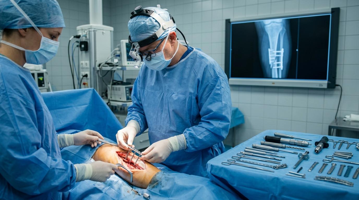
Emergency orthopaedic surgical suite with continuous fluoroscopy and intensive care monitoring.
Frequently Asked Questions About Arthroscopy
Injured Your Knee or Shoulder?
Consult Dr. Vishal Kasliwal for an accurate clinical assessment, MRI review, and personalized sports recovery plan.
Medically reviewed by: Dr. Vishal Kasliwal, M.S. Orthopaedics · Reg. No. MMC/2005/02/1098
Last updated: October 2026
Medical Disclaimer: This information is for educational purposes only and does not replace an individual medical consultation. Diagnosis, treatment options, and recovery timelines differ between patients.