
Hip Treatment & Hip Replacement in Nashik
Specialised clinical diagnosis and surgical care for hip pain, degenerative arthritis, and avascular necrosis (AVN)—from joint-preserving core decompression to precision ceramic total hip replacement. Evaluated and operated by Dr. Vishal Kasliwal at Kasliwal Joint Institute.
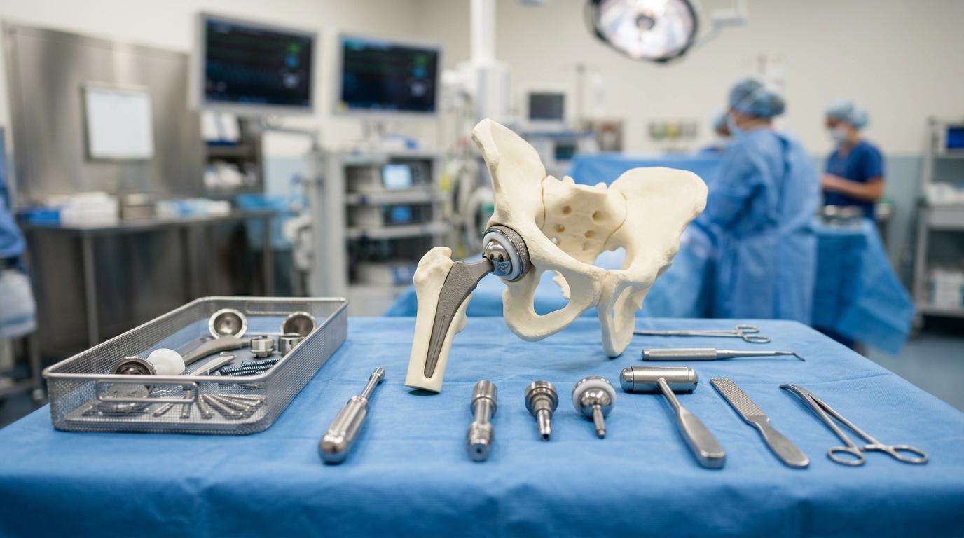
Common Hip Problems We Treat
Hip symptoms vary widely depending on the anatomical origin. Accurate clinical assessment with specialized standing pelvic imaging ensures targeted, conservative-first management.
Hip Osteoarthritis
Cartilage wear in the ball-and-socket joint causing groin stiffness, restricted bending, and limping during daily walking.
Avascular Necrosis (AVN)
Compromised micro-circulation to the femoral head ball, leading to bone ischemia, subchondral collapse, and severe pain.
Labral Tears & FAI
Cartilage rim tears and bone impingement causing sharp groin catching, pinching, and discomfort during hip rotation.
Inflammatory Arthritis (AS)
Systemic spondyloarthritis in younger adults causing progressive pelvic joint stiffness, fusion risk, and fixed flexion contractures.
Elderly Hip Fractures
Neck of femur and intertrochanteric fractures requiring prompt emergency surgical fixation or bipolar hemiarthroplasty.
Failing or Loose Hip Implant
Persistent pain, clicking, or loosening from previous surgery requiring specialist revision assessment.
Is the Pain from Your Hip or Your Back?
Differentiating true hip joint pathology from referred lumbar spine pain (sciatica) is essential for targeted, effective treatment.
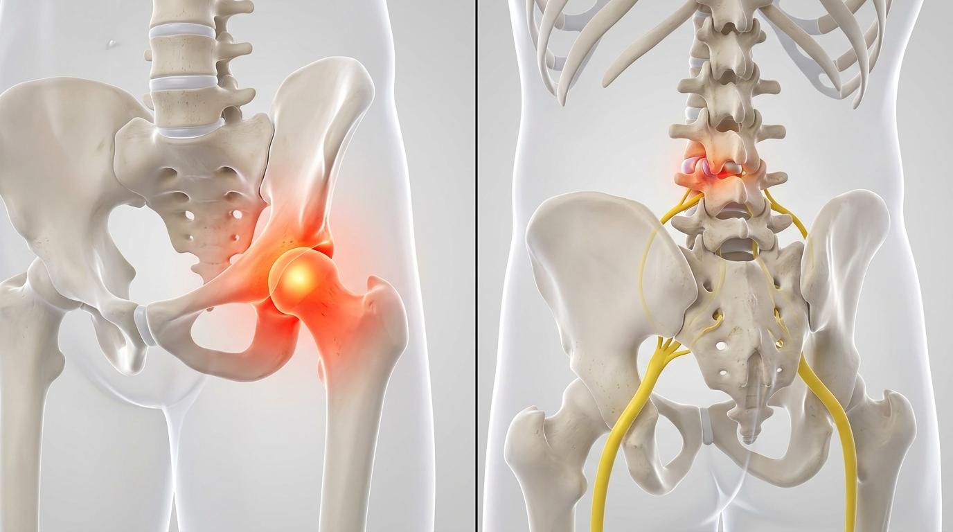
| Indicator | True Hip Joint | Lumbar Spine / Sciatica |
|---|---|---|
| Pain Location | Groin crease, front thigh, inner knee | Lower back, buttock, back of calf & foot |
| Pain Nature | Deep mechanical ache, stiffness, limp | Electric shooting, burning, numbness |
| Aggravated By | Walking, pivoting, car transfers | Prolonged sitting, coughing, forward bend |
| Key Daily Sign | Trouble clipping toenails or wearing socks | Pain shooting past knee into calf |
| Clinical Test | Positive FABER & FADIR hip stress tests | Positive straight-leg raise (SLR) test |
Hip Arthritis Treatment in Nashik
Hip arthritis is progressive wear of the smooth cartilage lining the ball-and-socket joint. We prioritize evidence-based conservative management to preserve your natural joint whenever possible.
Key Symptoms & Warning Signs
- •Deep groin or outer thigh pain aggravated by daily walking and weight-bearing
- •Stiffness making it difficult to put on socks, tie shoelaces, or clip toenails
- •A noticeable limp or sensation that one leg is becoming structurally shorter
- •Severe restriction in sitting cross-legged or getting down to the floor
- •Rest pain or nocturnal throbbing that disturbs sleep quality
Conservative Management Ladder First
Do You Need a Hip Replacement? Self-Assessment Checklist
Surgery is considered when arthritis pain severely limits daily life and non-surgical treatments stop working:
If you answered yes to 3 or more indicators, an objective surgical consultation is recommended.
Book Knee & Hip EvaluationAVN (Avascular Necrosis) of the Hip: Treatment in Nashik
Avascular necrosis (osteonecrosis) occurs when micro-circulation to the femoral head (ball) is compromised. Deprived of oxygenated blood, the bone tissue weakens and can collapse under daily body weight. Early detection via MRI is critical while the ball remains spherical and joint preservation is still possible.
Common Causes & Risk Factors for Hip AVN
Early Diagnostic Rule: Standard X-rays can appear normal in early AVN. An MRI scan of both hips is the gold standard for early detection when joint-preserving surgery is still achievable.
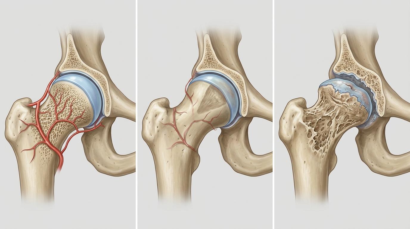
AVN Progression: Normal Blood Supply → Ischemia → Subchondral Collapse
| Stage | Anatomical Condition | Treatment Options |
|---|---|---|
| Stage 1–2 Pre-Collapse | Ischemia on MRI, but the femoral head retains its spherical round contour. | Joint Preservation: Core decompression & BMAC stem cell augmentation. |
| Stage 3 Crescent Sign | Subchondral bone fracture occurs; the femoral ball begins to flatten and lose sphericity. | Preservation is less predictable; replacement is considered based on symptoms and activity. |
| Stage 4 Joint Collapse | Total collapse of the femoral head with acetabular socket erosion and severe joint destruction. | Total Hip Replacement (THR): Reliably restores pain-free movement and equalizes leg length. |
Total Hip Replacement in Nashik
In a total hip replacement (THR), the arthritic femoral ball is replaced with a highly polished ceramic or metal head mounted on a titanium stem. The worn acetabular socket is lined with a biocompatible titanium shell and an ultra-durable crosslinked polyethylene or ceramic insert.
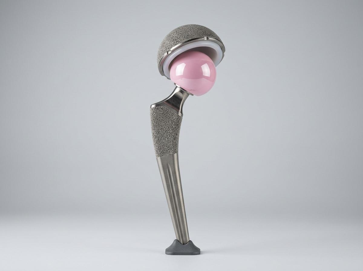
Uncemented Biological Ingrowth Total Hip Prosthesis
Digital Templating & Leg Length Equalization
Careful pre-operative digital planning determines the exact femoral stem offset, cup inclination angle (within the Lewinnek safe zone), and neck length to restore balanced leg lengths and stable soft-tissue tension.
Ceramic Bearings for Maximum Longevity
We utilize Delta ceramic femoral heads paired with highly crosslinked polyethylene or ceramic liners. These advanced bearing surfaces have microscopic wear rates, providing 15 to 20+ years of durability for active adults.
Tissue-Preserving Capsular Repair
Muscle-sparing surgical approaches with complete anatomical capsular reconstruction and external rotator repair minimize blood loss and virtually eliminate dislocation risk.
Step-by-Step Hip Replacement Journey
Pre-op Templating & Fitness
Cardiac, anaesthetic, and blood optimization with pelvic digital scanogram measurement.
Targeted Spinal Anaesthesia
Conducted under modern regional spinal anaesthesia with multimodal pain control for rapid awake recovery.
Surgical Procedure (1.5–2 Hrs)
Accurate cup reaming, stem insertion, trialing for full range of motion, and secure anatomical soft-tissue repair.
Day 1 Supported Walking
Guided mobilization with a walker begins within 24 hours under our physiotherapy team to stimulate circulation.
Stair & Transfer Training
Mastering chair transfers, bathroom independence, and stair navigation prior to hospital discharge.
Safe Home Discharge
Discharge home occurs within 2 to 3 days with complete wound care guidance and home exercise protocol.
Recovery Timeline After Hip Replacement
Because hip replacement relieves bone-on-bone pain immediately, rehabilitation progresses swiftly following a structured timeline:
Standing & First Steps
Supported standing and short corridor walking with a walker. Gentle ankle pumps and isometric gluteal contractions begin.
Independence & Stairs
Bed-to-chair transfers, independent toilet visits using a raised commode, and step climbing practice with our physiotherapy team.
Stick Walking & Dressing Review
Follow-up wound check; transition from walker to a single walking stick. Independent walking around the house.
Independent Walking
Most patients discontinue the walking stick and walk smoothly without a limp. Resumption of outdoor walks and light routines.
Driving & Office Work
Return to work, driving (once verified by clinical review), and domestic travel as hip precautions are gradually relaxed.
Full Active Life
Full return to swimming, brisk walking, golf, travel, and active routine with durable, pain-free hip mechanics.
Early Dislocation Precautions & Indian Home Setup
Surgical Safety & Risk Mitigation
Total hip replacement is an established and predictable surgery. We discuss potential risks openly and follow rigorous clinical protocols to minimize them:
Infection Control in Modular OTs
Surgeries are performed in positive-pressure laminar airflow operating theatres with HEPA filtration, strict sterile draping, and protocolized prophylactic antibiotics adhering to ICMR orthopaedic guidelines.
Blood Clot (DVT) Prevention
Deep vein thrombosis risks are mitigated with early Day 1 walking, graduated compression stockings, pneumatic calf pumps during bed rest, and supervised blood-thinning medications.
Dislocation Prevention Protocols
Using larger-diameter 32mm/36mm ceramic heads, accurate cup orientation, and anatomical posterior capsule/rotator repair significantly reduces dislocation risks to under 0.5%.
Leg Length Accuracy
Pre-operative scanograms and intraoperative caliper measurements are cross-checked before final component seating to restore balanced limb lengths and pelvic alignment.
Hip Fracture in the Elderly: Time-Sensitive Care in Nashik
A domestic slip or fall in an elderly family member resulting in a broken hip (femoral neck or intertrochanteric fracture) is a medical emergency. Prolonged bed rest in older adults rapidly triggers complications like chest infections, bed sores, and blood clots.
Prompt surgical intervention—ideally within 24 to 48 hours—allows early pain-free sitting and mobilization:
Painful, Loose, or Dislocating Hip Replacement?
If you had a hip replacement elsewhere that has developed persistent groin pain, clicking, loosening from bone, or recurrent dislocation, specialist revision assessment is available. We manage bone loss, component exchange, and dual-mobility stability solutions.
Hip Replacement Cost & Cashless Insurance in Nashik
Every surgical estimate is provided transparently in writing following your clinical consultation and pelvic X-ray review. The total cost is determined by four clinical factors:
Procedure Type
Core decompression vs total hip replacement (THR) vs bipolar hemiarthroplasty.
Bearing Surfaces
Choice of Delta ceramic-on-polyethylene vs ceramic-on-ceramic bearings.
Room Category
Choice of general healthcare ward, semi-private room, or private deluxe suite.
Stay Duration
Standard 2–3 day hospital stay or extended medical observation for co-morbidities.
Cashless Mediclaim Desk & TPA Empanelment
Our in-house insurance desk manages pre-authorization and documentation with all major public and private health insurance companies. We help verify your hip procedure coverage, waiting periods, and room category eligibility prior to admission.
Frequently Asked Questions About Hip Care
Hip pain, stiffness or difficulty walking?
Book an assessment with Dr. Vishal Kasliwal to diagnose the cause of your hip problem, evaluate your X-rays or MRI, and discuss the right treatment path.
Medically reviewed by: Dr. Vishal Kasliwal, M.S. Orthopaedics · Reg. No. MMC/2005/02/1098
Last updated: October 2026
Medical Disclaimer: This information is for educational purposes only and does not replace an individual medical consultation. Diagnosis, treatment options, and recovery timelines differ between patients.